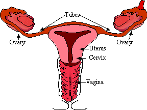
| Vagina | Cervix | Uterus | Ovaries | Fallopian Tube | The vagina extends from the vaginal opening to the cervix - the opening to the uterus. The vagina receives the penis during sexual intercourse, and is the birth canal through which the baby passes during labor. The average vaginal canal is three inches long, possibly four in women who have given birth. This may seem short in relation to the penis, but during sexual arousal the cervix will lift upwards and the fornix (see illustration) may extend upwards into the body as long as necessary to receive the penis. After intercourse, the contraction of the vagina will allow the cervix to rest inside the fornix, which in its relaxed state is a bowl-shaped fitting perfect for the pooling of semen. At either side of the vaginal opening are the Bartholin's glands, which produce small amounts of lubricating fluid, apparently to keep the inner labia moist during periods of sexual excitement. Further within are the hymen glands, which secrete lubricant for the length of the vaginal canal. The cervix is the opening to the uterus. It varies in diameter from 1 to 3 millimeters, depending upon the time in the menstrual cycle the measurement is taken. The cervix is sometimes plugged with cervical mucous to protect the cervix from infection; during ovulation, this mucous becomes a thin fluid to permit the passage of sperm. The uterus, or womb, is the main female internal reproductive organ. The inner lining of the uterus is called the endometrium, which grows and changes during the menstrual cycle to prepare to receive a fertilized egg, and sheds a layer at the end of every menstrual cycle if fertilization does not happen. The uterus is lined with powerful muscles to push the child out during labor. The ovaries are situated on the side of the uterus just below the
finger like projections of the fallopian tubes The
ovaries perform two main functions: production of female sex hormones -estrogen and
progesterone, and the production of mature ova, or eggs every month after puberty.
Of the two female hormones produced by the ovaries, estrogen is responsible for
development and maintenance of all secondary sex characters such as breast, shape of the
body, and maturity of the reproductive organs such as vagina, uterus, fallopian tubes, etc. Fallopian tube is the duct through which the egg must pass to reach the uterus. The tube is about 4" long and hang freely in the pelvic cavity. They are not directly connected to the ovaries but its end widens into a wide flower like opening that lies adjacent to the ovary. |